


-1675d2368c6531b4186d0c38e40719e5.png)


In ophthalmology, precision is everything. The ability to diagnose, treat, and monitor eye conditions confidently depends on how well clinicians can assess changes in the ocular structures over time. Yet, one of the most overlooked yet crucial tools in this process is photographic documentation.
A recent study published in the Cornea Journal, Controllable Errors in the Management of Infectious Keratitis by Todd P. Margolis and Anat Galor, highlights how diagnostic mistakes often stem from insufficient documentation. One of the key recommendations from the study is the importance of proper imaging to avoid misinterpretation and ensure accurate clinical decisions.

Accurate Diagnosis Through Imaging
High-quality imaging allows ophthalmologists to track the progression of conditions such as infectious keratitis, corneal ulcers, and other ocular pathologies. Without detailed photographic records, subtle details might be overlooked, leading to misdiagnoses or unnecessary treatments.
For example, infectious keratitis presents a significant diagnostic challenge. Patients often arrive late, sometimes already treated with corticosteroids or other agents, making it difficult to determine the exact cause of their condition. The study suggests that an accurate history, supported by detailed imaging, can mitigate errors and provide a clearer picture for decision-making.

The Link Between Poor Documentation and Malpractice Risks
Beyond diagnostic accuracy, lack of proper documentation is also a major legal concern for ophthalmologists. Studies show that insufficient or missing documentation is one of the most common reasons for malpractice lawsuits in the medical field. According to Healio, failure to record crucial details—including clinical images—can lead to disputes over whether a correct diagnosis and treatment plan were established.
Moreover, Ophthalmology Management emphasizes that even the best clinical decisions can be questioned if they are not properly documented. This reinforces the need for a systematic approach to medical imaging and record-keeping.
How a Data Management Platform Can Improve Outcomes
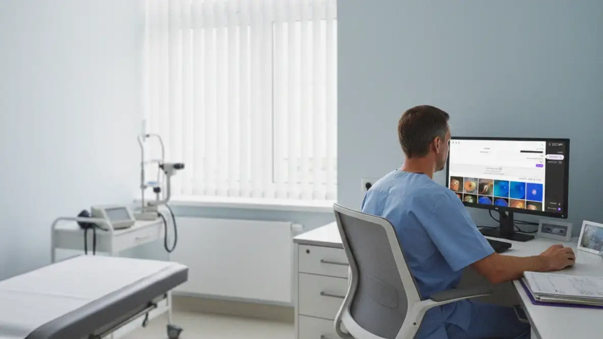
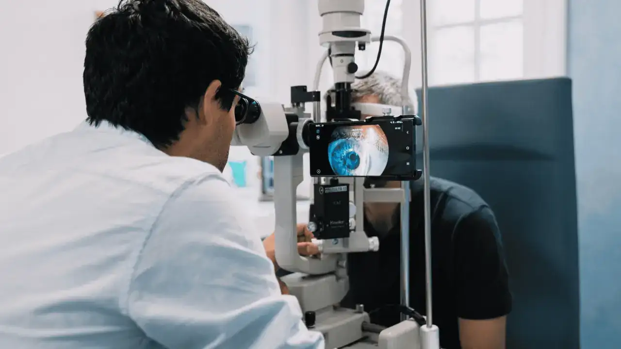
To ensure accuracy and reduce malpractice risks, ophthalmologists must integrate high-quality imaging with a structured data management system. A tool like MicroREC Connect allows clinicians to capture and store high-resolution images directly from their microscopes, keeping all relevant patient records in one accessible platform.
With a dedicated imaging and documentation system, practitioners can:
- Track disease progression with precise, high-quality images over time.
- Improve collaboration by sharing cases with colleagues or referring specialists.
- Protect themselves legally with comprehensive patient records.
- Enhance treatment outcomes by avoiding misdiagnoses and unnecessary procedures.

The Future of Ophthalmology: A Commitment to Documentation
The importance of detailed imaging and proper record-keeping cannot be overstated. In a field where precision defines success, having a structured documentation system is no longer optional—it is essential.
By incorporating advanced tools like MicroREC Connect, ophthalmologists can elevate their diagnostic accuracy, safeguard their practice, and ultimately provide the highest level of care for their patients. Investing in quality imaging and documentation today means better patient outcomes and fewer risks tomorrow.
Resources
- Margolis, T. P., & Galor, A. Controllable Errors in the Management of Infectious Keratitis. Cornea Journal, Volume 43, Number 2, February 2024.
- Healio – Lack of Documentation Most Common Reason for Malpractice Suits
- Ophthalmology Management – How to Avoid a Medical Malpractice Claim